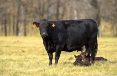

Findings from a recent Alberta project have implications for management practices that could help prevent the spread of pinkeye (infectious bovine keratoconjunctivitis, IBK) within a herd.
Relatively little attention has been given to pinning down the cause and prevention of pinkeye even though it is the most common eye disease of cattle worldwide, and highly contagious. The long-standing school of thought is that it is caused by Moraxella bovis bacteria and spread by flies feeding on eye secretions of infected cattle. Eye irritation from ultraviolet light, dust or abrasive forages is thought to predispose cattle to the infection by creating a point of entry for bacteria, explains Sarah Van Schothorst (nee Cottle), who tackled some new questions about this old disease while completing her bachelor of science degree in biology at the University of Calgary.
Read Also
Body condition, nutrition and vaccination for brood cows
One of the remarkable events of the past century related to ranching has been the genetic evolution of brood cows….
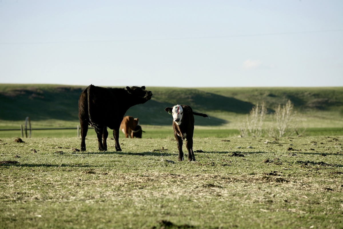
Van Schothorst is inclined to believe that bacteria must be transmitted in ways other than by flies because several herds in southern Alberta have histories of pinkeye occurring in winter and worsening throughout the summer, and many ranchers say that the period after weaning is a high-risk time for pinkeye.
“I had the idea when I looked at herds dealing with outbreaks that the eye and nose communicate with each other. In most mammals, the naso-lacrimal duct drains tears into the nose, therefore, perhaps aerosolized nasal secretions or even nose-to-nose contact would be the more likely predominant mode of transmission,” she explains.
Another perplexing aspect of pinkeye is that even though M. bovis is the only bacteria confirmed to cause pinkeye in cattle, it cannot be reliably isolated from the eyes of all infected cattle. Moraxella bovoculi, first identified as a distinct species in 2002, is becoming an increasingly common isolate in cultured samples from cattle with pinkeye. The role of M. bovoculi in the disease process isn’t clear because it has not yet been shown to reproduce the classical pinkeye lesions in the eye.
Van Schothorst’s project with Dr. Eugene Janzen, his assistant, Cynddae McGowan, and Dr. Claire Windeyer of the school’s faculty of veterinary medicine, investigated the frequency with which M. bovis and M. bovoculi occurred alone and together in the eyes and noses of calves with pinkeye during naturally occurring outbreaks. Samples were obtained by swabbing the eyes and noses of approximately 60 affected calves during outbreaks on seven ranches in southern Alberta.
M. bovis was found in 38 per cent of the eye samples and 10 per cent of the nose samples. M. bovoculi was found in 25 per cent of the eye samples and two per cent of the nose samples.
“Both species were cultured from samples from four of the seven ranches. The main point is that overall the same organism could be cultured from both the eye and nose of an individual 20 per cent of the time. We believe this supports the view that there is a similar microbiological biome in the eye and nose and, because of the naso-lacrimal duct communication, direct contact or aerosolized nasal secretions could be a potential route of pathogen transfer between cattle.
“So, we are moving away from the more traditional thinking of flies roaming across the eyes being the cause of transmission and that prevention should be more focused on handling management and perhaps feeding management. Not having as many cattle in the crowding tub at one time and spreading out feed so cattle don’t have to push around to eat will help minimize direct contact,” Van Schothorst explains.
She also wondered whether the bacteria are gaining resistance to oxytetracycline given that many ranchers treat pinkeye with intramuscular injections of long-acting oxytetracycline at the labelled dose and yet outbreaks continue in some herds. This is the standard treatment, especially when it’s not feasible to be handling cattle every day, although some producers prefer penicillin and other treatment options are available when affected cattle can be separated from the herd and treated daily.
Sixty eye swabs and 41 nasal swabs were sent to Prairie Diagnostics Services in Saskatoon to identify the bacteria involved and the susceptibility of M. bovis and M. bovoculi bacteria to all antibiotics commonly available to producers from their veterinarians. Some pooled samples were also sent to the Provincial Animal Health Laboratory at Guelph to test the isolates for the minimum inhibitory concentration (MIC) needed to kill the organism.
Using MICs as the measure of resistance, 20 per cent of the M. bovis bacteria were considered resistant to oxytetracycline at the labelled dose; however, none of the M. bovoculi showed resistance. M. bovis showed the highest level of resistance to penicillin, likely because it has traditionally been the drug of choice for treating pinkeye. Sensitivities of bacteria to antimicrobials will vary from operation to operation depending on the drugs used in the past.
“Knowing what kind of sensitivity these bacteria have toward the antimicrobials can really make or break whether your treatment method is effective. The best bet for producers who encounter outbreaks is to call a veterinarian. Getting a representative number of the calves sampled will tell you exactly what bacteria are present in the eye and can also determine the most effective method of treatment, which in the long run will save time and money,” Van Schothorst suggests.
Samples should be taken before treatment and early in the process when the eyes show excessive tearing and minimal visible lesions on the cornea. It is best to restrain cattle in a headgate rather than roping them because contamination from soil and other organisms in the sample make it difficult if not impossible to isolate the pathogens. The turnaround time for testing is two to three days.
More research is definitely needed to better understand the epidemiology of pinkeye and the pathogens involved to improve the routes and efficacy of treatments and vaccines.
Moraxella bovoculi update
An extensive study by Aaron M. Dickey et al. at the U.S. Meat Animal Research Center, Clay Center, Nebraska, investigating the genetics of M. bovoculi was published in February 2016 on the open-access website, BioMed Central.
Pinkeye progression

Pinkeye is highly contagious, but not life-threatening unless an animal is so severely affected it becomes blind in both eyes, leaving it prone to starvation, accidents and predation. That’s rarely the case and most of the time pinkeye clears up on its own, although the healing process can take up to five weeks.
The first signs are excessive tearing and extreme sensitivity to light. Extra blood vessels are visible toward the centre of the cornea to promote healing, which gives the white of the eye its telltale pink colour. The conjunctiva (inner layer of the eyelids) becomes red and inflamed and secretions become thick and yellowish. The surface of the eyeball turns cloudy and one or more ulcerated white spots develop on the cornea, often starting near the centre. A good indication that healing is underway is that the ulcer(s) begin to shrink. In some cases, the ulcer grows to cover the entire cornea before starting to heal. Severe infections that penetrate into the eyeball give it a yellow colour due to a buildup of pus inside. Permanent or partial blindness can result if ulcers rupture and eye fluids are lost, or if the inner tissue of the conjunctiva herniates and forms a bump over the eyeball.