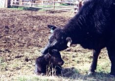

Environment, germs and immunity are top of mind when thinking of all the risk factors that could set the stage for pneumonia in cattle. The forgotten factor is one beyond producers’ control and the reason why pneumonia will always be a problem — anatomy.
Bovine lungs are very small relative to the animal’s oxygen requirements, explains Dr. Edouard Timsit, University of Calgary Veterinary Medicine and a veterinarian with Feedlot Health Management Services at Okotoks.
Read Also
Body condition, nutrition and vaccination for brood cows
One of the remarkable events of the past century related to ranching has been the genetic evolution of brood cows….
The total lung capacity of an adult cow is only 2.5 times greater than that of an average man, yet its resting oxygen requirement is more than 10 times greater. A cow’s lung capacity is 12 litres, its resting oxygen requirement is 124 litres per minute and it takes 30 breaths a minute to meet this demand. Compared to a species of similar size and structure, a horse’s lung capacity is 42 litres and its resting oxygen requirement is 49 litres per minute requiring only 11 breaths per minute.
The high airflow rate coupled with weaknesses of the bovine lung structure itself leave ways for bacteria, viruses and other contaminants to penetrate deep into the lungs where they trigger infection and inflammation.
The three categories of pneumonia from most-to-least prevalent overall are bronchopneumonia, interstitial pneumonia and embolic pneumonia. Timsit offers some pointers on how to tell one from the other along with suggestions for management practices that help outweigh the anatomical shortcoming.
Bronchopneumonia
Viral infections and other conditions that weaken the immune system or damage the mucosal lining of the respiratory tract can tip the scale in favour of bronchopneumonia, but it is bacteria that inflict severe damage. Several of the bacterial species involved in pneumonia are normally present in the upper airway where they do no harm. It’s only when stressors, such as weather, weaning, commingling and transportation, and/or viral infections knock down defences that bacteria can multiply in the nasal cavity and start spilling into the lungs to cause bronchopneumonia.
Don’t take a wait-and-see approach to treatment. Bacteria multiplying in the lungs sets a vicious cycle in motion that ends up with the immune system seemingly defeating its own purpose. Inflammatory substances released in the process of destroying bacteria draw a large amount of immune cells to the site of infection. The immune cells help clear infection early in the disease process, but they also release cellular contents that damage lung tissue. Lung tissue cells damaged by bacteria and the immune response are called lesions.
“Treat early when the bacterial load and lesions are mild,” Timsit says. “The major reason for treatment failure is the presence of lesions that are far too advanced for treatment. The basic foundations are to treat early enough and with the appropriate antimicrobial agent.”

Think “DART” — depressed, anorexic, respiratory changes, temperature — to catch pneumonia in the early stage. A depressed animal is usually away from the group, often with head and ears drooping, not showing much awareness of activity elsewhere and reluctant to move. Anorexia refers to weight loss or gauntness because the animal isn’t eating. Respiratory changes differ depending on the type and stage of pneumonia and could include changes in the rate and depth of respiration, nasal discharge and coughing. The general rule of thumb is to treat animals with temperatures above 104 F (40 C).
Mild depression and anorexia, with high fever, rapid, shallow breathing, a runny nose and cough are common in the early stages of viral pneumonia. The signs worsen if secondary bacterial pneumonia sets in. The cough usually sounds moist and breaths in and out become deep and laboured. Discharge from the nose and eyes thickens.
Early in the feeding period, even if no respiratory signs are evident, calves that are acting unusual and have a fever for no other apparent reason (lameness, diarrhea), are generally assumed to have bacterial bronchopneumonia, Timsit says.
The standard treatment is a three-day course of an antibiotic to slow or stop bacteria from multiplying. Prognosis is good with approximately 80 per cent recovering after the first treatment when given early enough. The case fatality rate can be up to 10 per cent.
In an outbreak, as many as 70 per cent of the animals in a pen could get sick and fatalities could add up quickly. Mass medication is worth considering because it will dramatically reduce the number of animals in a pen that get clinically sick, he adds. This control strategy is recommended especially if detection of sick animals isn’t optimal, that is a high case fatality rate because of high numbers of animals pulled too late.
Bronchopneumonia in feedlot cattle is most often seen during the 50 days after placement, ergo the old name, shipping fever.
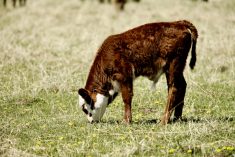
Pneumonia in sucking calves tends to increase from 70 to 150 days of age, which could be due to loss of herd immunity as maternal bodies wane, suggests a U.S. study involving 110,412 calves over 20 years. Another period of vulnerability identified in the study is from five to 30 days after birth, possibly due to poor passive immunity.
Nowadays, pneumonias of all types are generally referred to as bovine respiratory disease complex because the disease is the same even though it has many causes, Timsit explains.
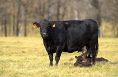
Reducing the risk factors starts at the cow-calf level by selecting females for calving ease and good udder and teat conformation, and providing proper nutrition so that calves are vigorous at birth and receive high-quality colostrum. Other basics include providing protection from the elements, preventing buildup of pathogens in the environment, and not mixing classes and ages of cattle. Vaccinating cows and calves can alter the risk factors, but can’t replace good management.
Risk factors for feedlot cattle can be difficult to eliminate because of a whole gamut of additional stressors that could include weaning, commingling, time in the marketing and transportation channels, dehydration and change of diet.
Timsit says giving an injectable antibiotic at processing on arrival to all high-risk calves has consistently been found to reduce sickness and death and improve performance. Vaccination on arrival coincides with a high-stress period and the immune response may not be adequate.
Aspiration pneumonia is a type of bronchopneumonia caused by breathing in foreign material, such as milk or medication due to improper drenching, amniotic fluid at birth, regurgitated rumen contents, or infectious material from lesions in the upper airway.
If the calf doesn’t die instantly, the telltale signs are sudden onset with severe symptoms usually accompanied by putrid breath because of the decaying material in the lungs.
It will probably be a rocky road to recovery for these calves involving a long run of antibiotics, pain medication and even a corticosteroid to relieve inflammation.
Interstitial pneumonia
Some known causes of interstitial pneumonia are viruses, migrating parasite larvae, chemical compounds converted from feed, allergic reactions, dust and spores from mouldy feed, and toxic gases (ammonia, smoke).
The cause of acute or atypical interstitial pneumonia (AIP) remains a mystery despite AIP being one of the most common pneumonias in feedlot cattle across Western Canada and the U.S.
“AIP is the most common pneumonia by far in Alberta feedlot cattle ready to slaughter,” Timsit says. “Some research suggests high tryptophan in the diet or a disruption in ruminal metabolism. All think it is associated with feeding, but there hasn’t been anything conclusive,” he says.

AIP usually comes on very late in the feeding period, frequently during hot, dry, dusty weather, and affects heifers more often than males, especially if heifers are receiving melengestrol acetate.
Signs of AIP that differentiate it from bronchopneumonia are the time frame in which it occurs (heavy cattle over 100 days on feed) and the sudden onset of laboured breathing with the neck stretched downward and an open, frothy mouth.
Timsit says the prognosis for recovery is guarded because there is slow or no response to routine therapy. An antimicrobial with short or no withdrawal time is usually given in case a secondary bacterial infection is involved, along with an anti-inflammatory drug. The best option in the long run might be to forego treatment and make arrangements for immediate salvage slaughter, especially if the animal has had bronchopneumonia in the past.
A second type of interstitial pneumonia is broncho-interstitial pneumonia (BIP). It is also known as upstairs-downstairs disease because it usually involves chronic bacterial bronchopneumonia affecting the lower front part of the lungs and interstitial pneumonia in the upper part extending to the back of the lung.
Death due to the interstitial pneumonia usually happens from 21 to 73 days after onset of bacterial bronchopneumonia. It is speculated that BIP is triggered by excessive inflammation and/or endotoxins released from gram-negative bacteria during the initial bronchopneumonia.
The next most common interstitial pneumonia is caused by an abrupt switch to lush pasture. Rumen bugs convert L-tryptophan in the forage into 3-methylindole, a chemical compound that is rapidly absorbed into the blood. Breakdown of the chemical in the lungs releases substances that damage lung tissues.
A good indication of interstitial pneumonia in pastured cattle is that it’s usually an outbreak situation. Signs in cattle that don’t drop dead immediately can range from severe to mild, with rapid, laboured breathing, loud grunts when air is expelled, mouth breathing and frothing at the mouth, but no cough. Coughing is frequent with pneumonia caused by parasites that also occurs in pastured cattle.
Most fatalities occur during the first two days and cattle that survive start to get better quite quickly after that with full recovery in about 10 days without treatment.
Timsit says the standard treatment is a non-steroidal anti-inflammatory drug, but this is one situation when the best treatment may be no treatment because any movement or excitement could be enough to cause instant death. Many animals recover on their own but the outcome for severely affected animals isn’t good because the lesions will probably be severe.
Prevention is the best medicine. Slowly adapt cattle to lush pasture over 10 days by feeding hay and supplement with monensin (slows the conversion of L-tryptophan), cut the pasture before turning cattle out or use it before it gets lush.
Embolic pneumonia
It usually starts with acidosis of the rumen, which triggers rumenitis that weakens the rumen wall allowing bacteria to penetrate into the bloodstream. Bacteria filtered out in the liver cause abscesses and if an abscess extends into the vena cava causing a thrombosis, small pieces off the thrombosis called emboli can end up in the bloodstream. Trapped in the tiny arteries of the lungs, emboli form abscesses resulting in an increase in blood pressure and weakening of the artery walls leading to aneurysms. Blood is released into the airway as the aneurysms rupture.
Bleeding from the mouth and nose is the definitive sign of embolic pneumonia. Affected animals become critical quickly, making treatment futile.
Embolic pneumonia can arise from emboli released from sites other than the vena cava, such as mastitis, arthritis or foot infections, but for the most part, managing feeding to prevent acidosis and liver abscesses will prevent embolic pneumonia.